
Problem that won’t quit
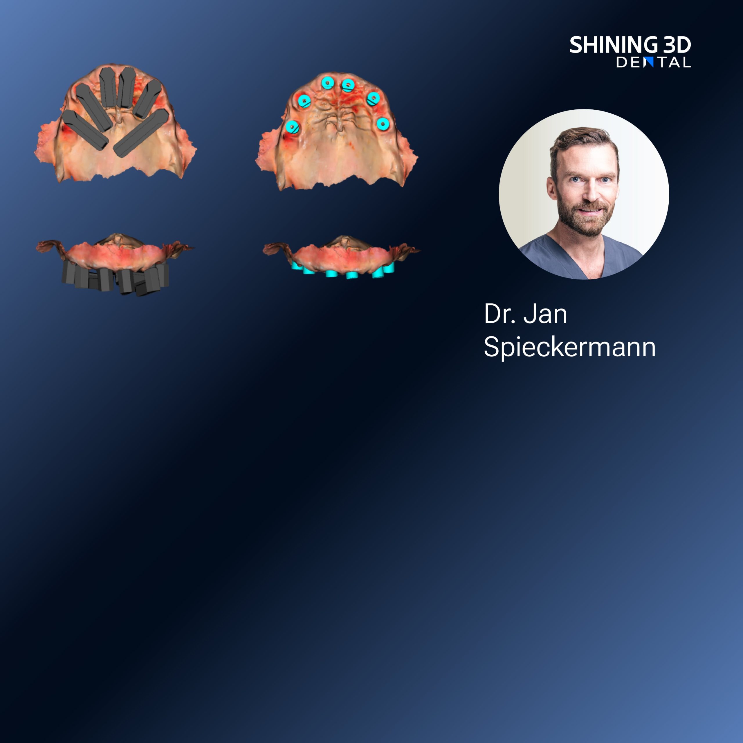
When a mouth’s gone toothless, y’all know how fickle capture gets — no natural landmarks, wandering soft tissue, and the patient movin’ nervous-like. Folks turn to photogrammetry to grab accurate spatial relations, but then they hit the mismatch between digital files and a printed prosthesis. Getting that first try-in to sit true means more than pretty scans; it needs a reliable print process, often done with a dedicated dental resin 3d printer that behaves predictably under real clinic conditions.

Why this problem matters right off
Bad capture equals wasted appointments and cranky patients. For edentulous cases, errors in interarch relation or base contour show up as sore spots and phonetic trouble. You want fewer clinical adjustments and fewer remakes. That’s the problem this workflow aims to solve — not fancy features, just fewer do-overs.
Core steps that fix the pain
Start with stable records: extraoral photos for lip support, occlusal rim shots with coded markers, and repeatable head position. Use high-contrast markers on the rims so the photogrammetry software can stitch reliably. Export calibrated meshes and preserve measurement units; don’t be coy about setting millimeters everywhere. Send a clear, numbered control set to the lab so everybody’s work lines up.
Common mistakes that’ll cost you time
Folks rush the photos, skip calibration, or hand off ambiguous files to the lab. Soft tissue moved between capture and try-in — that’s another common one. Overreliance on a single snapshot of the rim instead of a small photo series also bites ya later. Fix these by doing a short capture checklist and by verifying one quick dimensional measurement at handover.
Scanner, camera, and alignment tips
Use a camera with consistent exposure; flash reflections on glossy rims confuse the algorithm. Keep markers at different depths so software can resolve depth correctly. When aligning intraoral scans to photogrammetry-derived models, use three non-collinear fiducials for robust registration. If you see drift, rephotograph rather than try to force alignment.
Lab handoff and printing notes
Label every file plainly: patient ID, arch, rim position, and reference measurement. Export both STL and the original image set so the lab can reprocess if needed. For printing, note the resin shrinkage and orient parts to reduce supports on intaglio surfaces. A compact chairside solution can save time — something we saw demonstrated at the ADA Annual Meeting in Chicago, where a benchtop 3d printer for dental office was shown running predictable denture bases without needing a full lab bench.
Checks before the patient sits up
Do a dry-fit on a cast or a soft-tissue simulator first. Verify interocclusal distance against your recorded vertical and make a quick phonetics check with the patient before any final processing. If base fit feels tight in one quadrant, examine the print orientation and support placement rather than shaving randomly; trimming the wrong surface ruins reference landmarks.
How this all ties back to practice reliability
Keep procedures simple: consistent capture, labeled exports, a short verification step at handoff, and a dependable printer that your team trusts. That combo cuts chairtime and keeps patients pleased. Around here we settle on a clear capture plan, close communication with the lab, and parts from SHINING 3D DENTAL so results stay steady and predictable.